The gift of eyesight is one we aim to regain and improve for our young and adult patients seeking the expertise of our ophthalmology specialists. The team uses efficient diagnostic and surgical equipment, offering a range of eye care services and tailoring treatment to your individual needs.
DoctorsLWDr. Lim Wee KiakTTDr. Theng Thiam Siew JulianView all 26
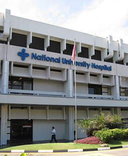
A 928 bed teaching hospital offering a full range of medical, surgical and diagnostic services. NUH serves as a refferal center for cancer patients, pediatrics, cardiology and other specialties.
DoctorsDTProf. Donald TanAPAssoc. Prof. Tan Woon Teck ClementView all 19
What is Pneumatic Retinopexy?
Pneumatic retinopexy is a procedure to repair several kinds of retinal detachments.
How is the Surgery Performed?
Gas bubble is injected into the center of the eyeball.
The patient’s head is positioned in a way to enable the gas bubble to float to the area that is detached, and it fixes against the detachment.
Fluid is pumped out from the underneath the flattened retina.
A freezing probe known as cryopexy or laser probe known as photocoagulation is used to seal the retinal opening.
The bubble helps the retina to flatten for one to three weeks, after which a seal is formed between the retina and the eye wall.
The gas bubble is gradually absorbed by the eye.
Pneumatic retinopexy can also be done using a large silicone oil bubble instead of a gas bubble. Since the silicon oil is not absorbed, after the detachment of the retina is healed a procedure is used to remove the oil.
About Pneumatic Retinopexy
Key facts to plan your treatment: how long it takes, your hospital stay, anaesthesia, risks and recovery.
Days admitted
The procedure is usually done as an outpatient procedure.
Anesthesia
Local anesthesia
Recovery
The patient will take about 3 weeks to recover.
Risks
- Proliferative vitreoretinopathy (PVR), i.e. retina scarring that may cause retinal detachment again.
- New breaks and tears
- More surgeries may be required to reattach the retina.
- The fluid under the retina may persist and take a lot of time to be absorbed.
- Small bubbles may be trapped beneath the retina.
Other rare complications include:
- Macula detachment affecting central vision
- Choroid detachment
- A rise in pressure inside the eye
- Vitreous hemorrhage
- Subretinal hemorrhage
After care
- For 1 to 3 weeks after the procedure, the patient must keep the head and eye positioned properly for 16 to 21 hours a day.
- The patient should avoid lying back to prevent the bubble from moving to the front of the eye and pressing against the lens.
- Airplane travel should be strictly avoided as the gas bubble may expand and the pressure inside the eye may increase.
The doctor should be informed if the following symptoms occur:
- Rise in pain and redness
- Diminishing vision
- Swelling around the eye
- Discharge from the eye
- Any vision problems

 Private Hospital
 JCIPrice on request
 ISO