Mitera hospital is a specializes hospital for women and children, with comprehensive pediatric, gynecological and maternal healthcare services. It is affiliated with Harvard’s Mass General Hospital for Children and Yale Children’s Hospital.
This is a subspecialty of radiology which involves imaging of children. It takes into account that organs follow growth patterns and phases in a growing body from infants to adolescents.
Who is a Pediatric Radiologist?
This is a specialist who uses imaging techniques and equipment t diagnoses diseases and injuries in infants, children, and adolescents.
Training
Pediatric radiologists have a degree from a medical school, at least one year of training in clinical medicine, four years training in diagnostic radiology, at least one year of training in the diagnosis of children using imaging equipment and additional years of training in the diagnosis of infants and children using imaging equipment.
What Do Pediatric Radiologists Do?
Pediatric c radiologists expertly select the best imaging technique to diagnose medical and surgical problems in children. Imaging techniques used in pediatric radiology include ultrasound d, x-ray, nuclear medicine, computed tomography (CT) and magnetic resonance imaging (MRI).
They ensure that the test is accurately and safely performed, interpret the results and make the appropriate diagnosis.
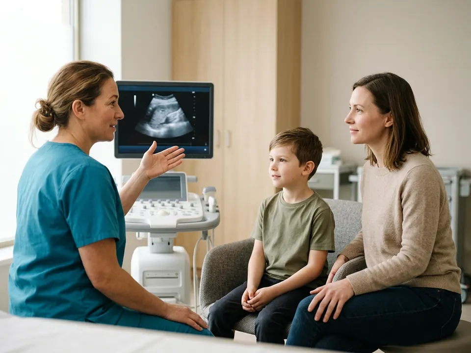
Pediatric radiologists have to be especially patient and friendly to help gain the child’s trust so as to perform the tests. Most will wear colorful uniforms and decorate their offices and equipment rooms with pictures that children like. They also use equipment designed for children which make the test more comfortable for the children and exposes them to minimal radiation.
Pediatric illnesses that require imaging include:
Leukemia
Osteosarcoma
Meningitis
Wilms' tumor
Infant respiratory distress syndrome
Teratoma
Juvenile idiopathic arthritis
Congenital abnormalities
Greenstick fractures
Cardiovascular conditions
Urologic conditions
Chest and respiratory conditions
Gastrointestinal conditions
Skeletal conditions
Neurological conditions
Neonatal conditions
Orthopedic oncology conditions
Orthopedic conditions
Oncology conditions
Traumatic injury
Imaging Technologies
Radiography and x-rays: These use electromagnetic radiation to view the body’s internal structure by creating images of the inside of the body. The images are usually in shades of black and white.
Ultrasound: This uses high-frequency waves to create images of the inside of the body. During the procedure, the image is displayed on a monitor.
Magnetic Resonance Imaging (MRI): This method uses radio waves and a magnetic field to create images of tissue and organs inside your body.
Computerized Tomography (CT or CAT scan): This imaging technique examines inside the body and displays the anatomical details of blood vessels very clearly. A contrast agent may be used for clearer images.
Nuclear Imaging (SPECT): It provides high-resolution images that can be represented as cross-sectional slices in three dimensions.
Positron emission technology (PET/CT) Scan: This combines CT and PET in one machine to produce three dimension images that are very clear.
Before the procedure
If the child is old enough to understand you can explain to them why the test is being done to reduce their anxiety.
You can practice the positions they will be in during the procedure and tell them how the test will feel and describe the equipment that will be used.
You can suggest ways of staying calm such as counting and breathing deeply.
After the procedure
After the child has completed the tests your doctor will discuss with you the results and diagnosis and make an appropriate treatment plan.
About Pediatric Radiology
Key facts to plan your treatment: how long it takes, your hospital stay, anaesthesia, risks and recovery.
Recovery
Information coming soon. Request a consultation for personalized recovery guidance. Get Quote →
After care
Information coming soon. Request a consultation for personalized recovery guidance. Get Quote →
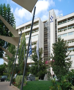 Women & Children's Hospital
 ISOPrice on request